


Research Highlights
PI McGlynn and colleagues develop new microscopy approaches to learn about "wild" cells.
PI McGlynn and colleagues develop new microscopy approaches to learn about "wild" cells.
While our knowledge of biological diversity is rapidly expanding thanks to improvements in sequencing and analysis techniques, the fast majority of cell types on the planet have never even been seen. We don't know their shape, size, physical nature of their interior, nor about their physical surroundings. To expand what is possible to be seen, we developed new microscopy techniques that are suitable for looking at cells from natural, non-laboratory, "wild" environments.
The goal was to link phylogenetic information - the type that can be gained through sequencing operations - to the physical nature of these "wild" and uncultivated cells. McGlynn and collaborators linked common forms of microscopy that are usually not performed in tandem, to be able to link the phylogeny and fine structure of the cell. Building off of that ability, the team developed ways of visualizing cell fine structure (nanometer resolution) in 3D. Surprisingly, unique physical properties of closely related cells could be seen, which could not be easily distinguished from a purely genomic analyses. With these new tools, it is possible to study cells from the wild in greater detail than before, towards the ultimate goal of linking form, function, and evolution, as we expand our awareness of microbial biodiversity. Of course these techniques can also be applied to the study of protocells - and like the cells reported on in this paper, they may have some interesting things waiting to be discovered in them; if we just look!
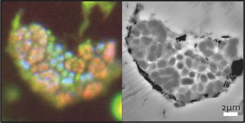
On the left, an epifluorescence image after undergoing fluorescence in situ hybridization (FISH) with specific archaeal and bacterial probes which color cells based on their phylogeny. On the right, a transmission electron microscopy (EM) image of the same cells. Together the techniques are termed by McGlynn and colleagues as FISH-EM.
Note and citation:
McGlynn and colleagues work, which makes use of epifluorescence and electron microscopy techniques, was published in the Journal Applied and Environmental Microbiology and was highlighted by the Journal Editors as a article of particular interest.
| Journal: |
Applied and Environmental Microbiology (84:e00399-18) |
| Title of original paper |
Subgroup characteristics of marine methane-oxidizing ANME-2 archaea and their syntrophic partners revealed by integrated multimodal analytical microscopy |
| Authors: |
Shawn E. McGlynn1,2,5, Grayson L. Chadwick1, Ariel O'Neill1, Mason Mackey3, Andrea Thor3, Thomas J. Deerinck3, Mark H. Ellisman3,4 and Victoria J. Orphan1 |
| Affiliations |
1Division of Geological and Planetary Sciences, California Institute of Technology, M.C. Pasadena, CA 91125, USA 2Biofunctional Catalyst Research Team, RIKEN Center for Sustainable Resource Science (CSRS), Wako City, Saitama 351-0198, Japan 3National Center of Microscopy and Imaging Research (NCMIR), Center for Research on Biological Systems, University of California, San Diego (UCSD) School of Medicine, La Jolla, CA 92093-0608 4Department of Neurosciences, UCSD La Jolla CA, 92093-0608; Salk Institute for Biological Sciences, La Jolla, CA; and HHMI Janelia Research Campus, Ashburn, VA. 5Earth-Life Science Institute, Tokyo Institute of Technology, Ookayama, Tokyo, 152-8550, Japan |
| DOI | 10.1128/AEM.00399-18 |
ELSI Researchers|Shawn McGlynn